Clinic Consultation in Kelowna / BC, excels in providing Ultrasound exam services, utilizing high-frequency sound waves to create real-time images of the body's internal organs, blood flow, and tissues. This non-invasive diagnostic tool is essential for a wide range of medical evaluations, including pregnancy monitoring, abdominal organ assessment, and detection of soft tissue conditions. Our clinic is equipped with the latest ultrasound technology, operated by certified sonographers and supervised by experienced radiologists, ensuring high-quality imaging and accurate diagnoses. Clinic Consultation prioritizes patient comfort and safety, offering a serene environment for all ultrasound procedures. Our healthcare professionals are dedicated to guiding patients through the preparation process, conducting the exams with utmost care, and providing detailed explanations of the findings. For individuals in Kelowna / BC, seeking reliable and comprehensive Ultrasound exam services, Clinic Consultation is the preferred destination, renowned for our commitment to excellence in patient care and diagnostic accuracy.
Ultrasound in Kelowna / BC
Ultrasound in Kelowna / BC
Ultrasound, also known as ultrasonography, is a diagnostic imaging technique that uses high-frequency sound waves to create real-time images of the body’s internal organs and tissues. It is a non-invasive, safe procedure commonly used in medical fields such as obstetrics, cardiology, and general diagnostics. Unlike other imaging methods, ultrasound does not use radiation, making it suitable for pregnant women and children.
What is Ultrasound Used For?
Ultrasound is used to diagnose and monitor a wide variety of medical conditions. It is commonly employed to monitor fetal development during pregnancy, assess organ function, detect abnormalities such as tumors or cysts, and guide certain medical procedures like biopsies. Its versatility makes it an essential tool in fields like obstetrics, cardiology, and abdominal imaging. Additionally, it is used in emergency medicine to assess trauma or internal bleeding.
How Does Ultrasound Work?
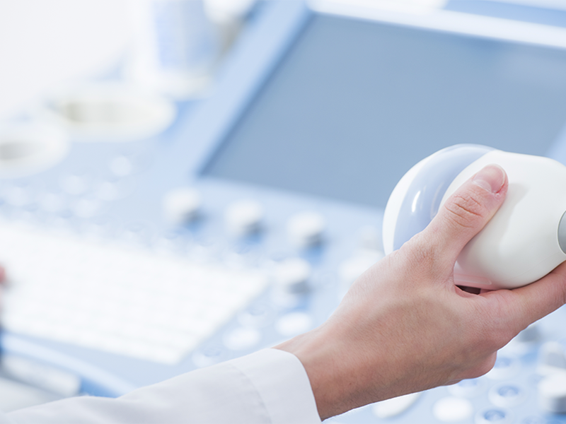
Ultrasound works by emitting sound waves through a handheld device called a transducer. These sound waves travel through the body, bouncing off tissues and organs. The returning echoes are captured by the transducer and converted into real-time images on a monitor. To improve sound wave transmission, a gel is applied to the skin where the transducer is placed. This method allows for a safe and painless examination of internal structures.
What Are the Types of Ultrasound?
Abdominal Ultrasound: This is used to examine organs like the liver, gallbladder, pancreas, and kidneys. It helps detect abnormalities such as stones, tumors, or infections.
Obstetric Ultrasound: Primarily used in pregnancy to monitor the fetus, assess development, and identify any potential complications, such as positioning or placental issues.
Doppler Ultrasound: This specialized ultrasound evaluates blood flow in arteries and veins. It is used to detect blockages, clots, or narrowing of blood vessels.
Transvaginal Ultrasound: This involves inserting a transducer into the vagina to get clearer images of the female reproductive organs, including the uterus and ovaries.
Breast Ultrasound: This is used to detect lumps or abnormalities in the breast tissue and is often a complement to mammography, especially in women with dense breast tissue.
What Diseases Can Be Detected by Ultrasound?
Ultrasound can detect numerous diseases and conditions. In the abdominal region, it can identify gallstones, liver tumors, kidney cysts, and pancreatic inflammation. In obstetrics, it helps diagnose fetal abnormalities and monitor pregnancy health. The Doppler ultrasound is essential in identifying vascular diseases such as deep vein thrombosis and aortic aneurysms. For cardiac health, it can detect valve defects and other structural abnormalities in the heart.
When is Ultrasound Indicated?
Ultrasound is indicated when a doctor needs to evaluate organs, tissues, or blood flow. It is routinely used during pregnancy and is essential for evaluating symptoms like abdominal pain, swelling, or unexplained weight loss. It is also used in emergency situations to detect internal bleeding or organ damage and can be an initial diagnostic tool for suspected tumors, cysts, or infections.
Pre- and Post-Care for Ultrasound
For most ultrasounds, minimal preparation is required. For an abdominal ultrasound, patients may be asked to fast for several hours to reduce gas that could obscure images. For pelvic ultrasounds, a full bladder is often needed to improve visualization of the pelvic organs. After the procedure, there are no specific post-examination instructions, and patients can typically resume their daily activities immediately.
Contraindications for Ultrasound
Ultrasound is a highly safe procedure with virtually no contraindications. Since it does not involve radiation, it is suitable for pregnant women and individuals of all ages. However, in cases of open wounds or severe skin conditions, it may be necessary to avoid scanning those areas to prevent discomfort or infection. Additionally, ultrasound might be less effective in imaging deep organs in patients with obesity.
Alternatives for Those Who Cannot Undergo Ultrasound
For patients who cannot undergo ultrasound or in cases where ultrasound cannot provide sufficient detail, alternatives such as CT scans or MRI are available. CT scans are often used for more detailed imaging of bones and tissues, while MRI is ideal for soft tissue evaluation, such as the brain or muscles. Both provide high-resolution images and can complement or substitute ultrasound when needed.
This examination is available at Clinic Consultation, where you can schedule your ultrasound appointment with experienced professionals for accurate and detailed diagnostic results.
Click the button below to schedule your appointment online.